Syringe pump technique for cell infusion into the fetal posterior kidney.
Takafumi Kuroda1, Shuichiro Yamanaka1, Kei Matsumoto1, SHI Chenyao2, Masashi Ikeuchi2, Takashi Yokoo1.
1Internal Medicine of Nephrology and Hypertension, The JIkei University, Tokyo, Japan; 2Department of Precision Biomedical Engineering, Institute of Science Tokyo, Tokyo, Japan
Background: End-stage renal failure is a disease requiring renal replacement therapy, and the only fundamental treatment is renal transplantation. However, due to the current shortage of donors, hemodialysis is the main method of treatment.
Xenotransplantation is attracting attention as a solution to the shortage of donors. However, xenotransplantation uses mature kidneys and rejection must be controlled. Regenerative medicine is attracting attention as another possibility. There are many challenges in creating mature organs with function from iPS cells.
For this reason, we are developing a technique for transplanting organ-specific progenitor cells into patients by transplanting the cells into a heterologous body, using a heterologous fetal organ as a scaffold for maturing organ-specific progenitor cells in vivo. This constitutes a broad form of foetal organ complementation, attempting organ regeneration by borrowing the developmental programme of xenogeneic animals.
Conventional manual transplantation techniques were challenging due to issues such as hand tremors, difficulty in fine depth adjustment, and the lack of reproducibility. Additionally, since the surgeon's breath was used to expel the cells, there were concerns regarding cleanliness.
Objective: In this study, we aimed to stabilise puncture and improve cleanliness by using devices when injecting cells into the posterior kidney. We also verified whether the injected renal progenitor cells colonised and differentiated in the posterior kidney or became chimeric, and whether the hind kidney itself developed after transplantation, using organ culture.
Methods: Cells from the fetal posterior kidney of C57BL/6JJmsSlc mice were used for transplantation. The cell solution was aspirated into a glass tube and mounted on a syringe pump with a valve mechanism.
14 days old mice foetuses were used for transplantation. We punctured the posterior kidney and injected cells under a fluorescence microscope. The injected cells were observed under a fluorescence microscope and assessed by immunostaining. The transplanted posterior kidney was also transplanted into male mice and development was checked.
Results: Using a syringe pump, we injected cells into the posterior kidney of mice and observed that the fluorescently labelled transplanted cells spread under the hindkidney capsule. Furthermore, even after peeling the posterior kidney from the foetus, we confirmed that the cells were injected into the hindkidney without damage to the capsule or cell leakage.
Immunohistochemical analysis revealed that the fluorescently labelled cells differentiated into renal tissue beneath the renal capsule. Chimeric formation with some self-tissue was also observed.
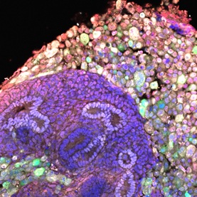
Furthermore, after transplanting the posterior kidney into mice following injection of renal progenitor cells, we confirmed renal enlargement and glomerular development in the transplanted posterior kidney when it was recovered two weeks later.
References:
[1] Cell transplantation
[2] fetal organ complementation
Lectures by Takafumi Kuroda
| When | Session | Talk Title | Room |
|---|---|---|---|
|
Thu-23 18:30 - 19:30 |
Poster Session | Syringe pump technique for cell infusion into the fetal posterior kidney. | Hall A2-A4 |